We’ve all seen and marveled at them: perfect fossils of gargantuan dinosaurs or other exotic creatures from the ancient world. But the truth is, sometimes there’s more than meets the eye. From signs of soft tissue and the remains of million-year-old meals, to delicate fossilized anatomy lost in the surrounding rock, to paint and plaster artfully applied by human preparators – many fossils have scientifically important features that are hiding in plain sight.
A research associate at The University of Texas at Austin is pioneering a new method for bringing these overlooked treasures into vivid detail. Called progressive photonics, the technique involves shooting photos of specimens under a range of lighting environments. The goal of the technique is to broaden the kind of information that can be extracted from fossils by diversifying and improving photo documentation.
The results can be stunning.
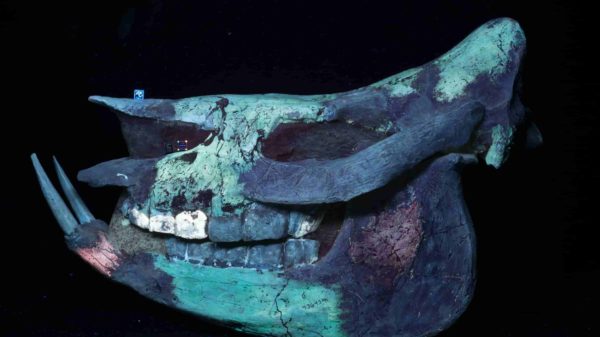
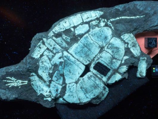
SCULPTED PAST: UVC light reveals that this Asian rhino skull is made up of a mosaic of material – so much that it has more in common with a work of art than a skeleton. The skull includes bones from at least two other sources (pink areas), which the scientists suspect came from unknown fossil specimens. The dull, dark purple is plaster, paint or adhesive. There is some original bone: the glowing green portions on the top of the specimen, mouth and bottom jaw.
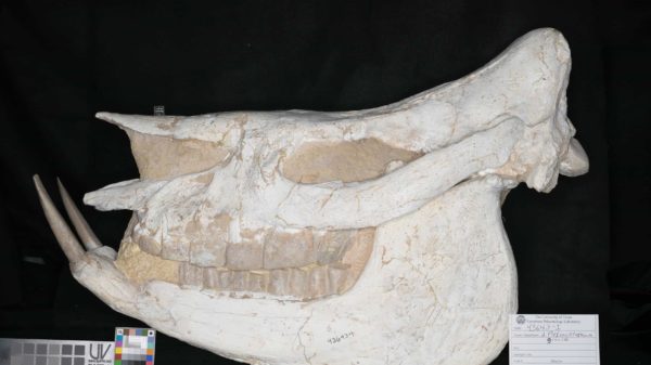
HIDING IN PLAIN SIGHT: The splashes of color that appear under UV light are nowhere to be seen under normal lighting. Bone and plaster blend perfectly together – making for a pretty skull, but a lousy scientific specimen.
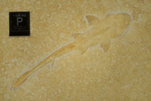
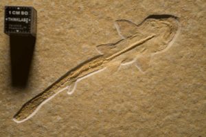
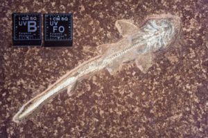
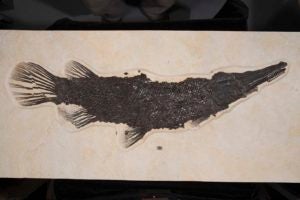
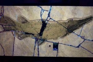
SHINE A LIGHT: Putting fossils under new lights offers new views. No one knew that this tiny fossilized shark, only a few centimeters long, had its gill anatomy preserved until it was examined under UVB illumination (right). In turn, a picture taken under polarized light (left) shows the outline of the body and vertebrae, which is further enhanced when the shark is viewed under oblique visible light (middle).